
Ex-Vivo MicroCT Scanning Facility
Instrument Details
The Bruker Skyscan 1272 offers high-resolution X-ray tomography with isotropic resolution from 1 to 50 microns, a 16Mpixel CMOS camera, and an adjustable stage. It supports fields of view up to 10 mm (1 micron) or 70 mm (50 microns) with triple offset scanning.
It excels in 3D imaging of mineralized tissues and can also image soft tissues with contrast agents (e.g., IKI, Lugol's, PTA). Vascular networks are visualized using perfusion fixation with agents like MicroFil. An automated changer enables batch imaging for up to 16 samples.
Data can be processed using 3D Slicer via the SlicerMorph extension or on MorphoCloud. In-house workstations are preloaded with 3D Slicer and SlicerMorph, and optional cloud-based processing is available through MorphoCloud.
Usage
NIH-funded investigators access the instrument for free, while training is required for self-use. Fee-based services are available.
Contact, Scheduling, and Additional Resources
Primary contact for details: Dr. Murat Maga
Calendar availability and additional resources
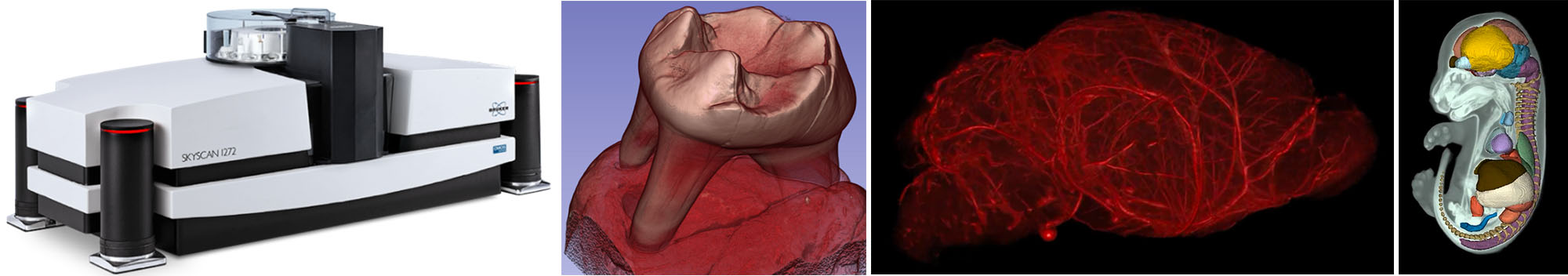
Pictured (left to right): The Bruker Skyscan 1272; A 3D rendering of a mammalian molar; A 3D vascular network in an adult mouse brain; A contrast enhanced CT of an E15 mouse fetus and its segmentation