Glue Embolization a Game-Changer in Treating Venous Malformations in Extremities: A Q&A With Drs. Giri Shivaram, Antoinette Lindberg and Eric Monroe
March 1, 2016
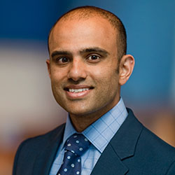
 Dr. Giri Shivaram
In 2013, members of the Vascular Anomalies team at Seattle Children’s developed a method to use a medical version of super glue to treat venous malformations in the head and neck area. This glue embolization process has been highly successful in removing malformations altogether.
After seeing how well the process worked, interventional radiologists Drs. Giri Shivaram and Eric Monroe and orthopedic surgeon Dr. Antoinette Lindberg decided to try using it to treat malformations in extremities.
While sclerotherapy has been the standard of care for most of these malformations, it’s a painful treatment that must often be repeated several times. Glue embolization enables a malformation to be surgically removed in one visit with much better results. The procedure is less painful than sclerotherapy and has a faster recovery time.
How can venous malformations affect a child’s quality of life?
Shivaram: Venous malformations
may be very small and not cause any problems, or quite extensive and, in some cases, encompass almost the entire leg or arm. They can cause a lot of pain and disability. While they’re not tumors, they can result in a child not being able to walk or use their arm.
What methods are used to treat venous malformations?
 Dr. Antoinette Lindberg
Shivaram: Historically, there hasn’t been a great way to treat them. In interventional radiology (IR) we would use sclerotherapy, which involved injecting the venous malformation with medications to scar the veins down, but this often required multiple sessions with variable results. Surgical removal alone was problematic because malformations bleed a lot and it’s hard to get around them.
This new hybrid procedure has revolutionized the way they are managed. While the patient is under anesthesia, we inject glue into the channels of the venous malformation, which we can see with ultrasound. The patient then goes to the operating room (OR) where the surgeon can see the malformation (due to the hardened glue) and surgically remove it. There is much less bleeding since the vascular channels of the malformation are blocked. We have performed more than 40 of these procedures over the last year and our patients have done amazingly well.
How long does it take from the time the glue is injected into the venous malformation until it can be surgically removed?
Lindberg: Basically, the time it takes to move them from the IR suite, where the injection is done, to the OR.
Shivaram: The glue we use is a cyanoacrylate glue, which is similar to household Krazy Glue. We inject it into the veins and it sets almost instantly to form something like a die-cast mold of the malformation. The amazing thing is that we can do it under one anesthetic. If you do sclerotherapy, kids have to come in for multiple treatments, each separated by a few months and under general anesthesia every time. We’re combining everything into a single-stage procedure.
How does removal using glue embolization stack up against standard surgical removal?
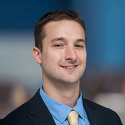
 Dr. Eric Monroe
Lindberg: Surgical removal of malformations was notoriously difficult. They bleed quite a bit and, unlike tumors, they’re not a distinct object where you can clearly identify its edges. On MRI scans, malformations are sometimes very distinct, but once you get in there it’s difficult even for the best surgeon to identify where the malformation starts and stops. Also, recurrence rates were 50% or higher, so many people chose to not even try to remove them.
It looks like Seattle Children’s and Boston Children’s are the only places where this kind of glue embolization is performed. Why aren’t there more?
Shivaram: It’s really hard to get the right balance of people. We’re lucky that we have a multidisciplinary vascular anomalies program here, led by Dr. Jonathan Perkins. Seattle Children’s has the surgeons and the interventionalists in one place, which makes it easy to coordinate the procedure and the surgery. We also have created a very organized method for intake and imaging of these patients.
What’s the recovery like?
Lindberg: It’s highly variable, depending on the location of the venous malformation. Some are more superficial and patients recover faster. Those that are deeper and involve more muscle take a little longer to heal. We don’t usually have to put very many restrictions on patients after the procedure. For example, if we remove a malformation from their leg, patients can usually walk on it right away.
Monroe: When malformations are found early, they’re relatively easy to deal with using this technique. But if left untreated, and if alternative or older therapies are used, management becomes more challenging on our end. Getting the word out will help us catch a lot of these earlier and have even better outcomes.
What is most important for providers to know about this procedure?
Lindberg: Send patients to see us early so we can start following them, even if their symptoms aren’t too bad. Patients and their family members need to know what they’re dealing with and what are their options.
Shivaram: Getting the correct diagnosis and developing a treatment plan with the family is also key, so other potentially harmful therapies are avoided. Even if we’re not going to treat them immediately, we want to avoid misdiagnosis and inappropriate treatment. When you have a procedure that works, you want to offer it to the children who need it. If I was a parent of a child with a venous malformation suitable for glue embolization surgery, I would definitely want them to get this treatment rather than sclerotherapy.
What indicators does a provider need to look for and when should they refer a patient? Does the referral need to come through interventional radiology?
Shivaram: An indication for referral would be a symptomatic mass or lesion thought to be a vascular anomaly, such as a venous or lymphatic malformation. A patient with asymptomatic lesions should also be referred to us so we can determine the need and timing of any later intervention. Referrals can come through either interventional radiology or orthopedic surgery. Referrals for orthopedic surgery should be directed to Dr. Lindberg.