How 3D Printing is Advancing Pediatric Surgical Care in the Pacific Northwest
May 7, 2025
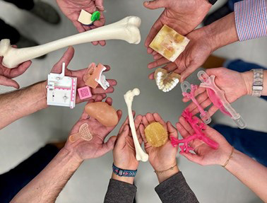 Members of Seattle Children’s Custom Care team hold some of their 3D-printed creations.
What if a piece of plastic could help improve the outcome of a patient’s orthopedic surgery? Thanks to the collaboration between Seattle Children’s Orthopedics and Sports Medicine and Custom Care teams at Seattle Children’s, it’s possible.
In partnership with Seattle Children’s Continuous Improvement and Innovation and Advanced Surgical and Procedural Innovation, Research and Education programs, the Custom Care team creates digital and 3D-printed models of complex surgical patients across the hospital, including for many orthopedic patients. These digital and life-size physical models provide unique training opportunities for surgical faculty, residents and fellows to practice procedures and hold critical discussions before patients undergo complex surgeries.
In the Pacific Northwest, Seattle Children’s leads the way in developing these cutting-edge pediatric patient models — accelerating innovation in surgical planning and contributing to life-changing research.
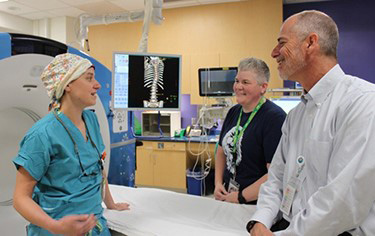 From left to right: Dr. Bauer meets with Jamie Ryan, computed tomography (CT) technologist, and Burt Yaszay, MD, chief and medical director, Orthopedics and Sports Medicine, to review a CT scan of a patient’s spine. These state-of-the-art X-Ray images provide multidimensional, detailed pictures of body tissues and structures.
Seth Friedman, PhD, principal investigator in the Center for Respiratory Biology and Therapeutics and manager of innovation imaging and simulation modeling, oversees the program. He calls his team an “equal-opportunity shop” for surgical teams across Seattle Children’s, serving Orthopedics and Sports Medicine, Otolaryngology, Dental, Neurosurgery, Cardiology, and Craniofacial Departments, to name a few. From tiny airways to intricate spines, the Custom Care team helps improve outcomes for patients across a broad range of specialties.
Jennifer Bauer, MD, head of Seattle Children’s Spine Surgery Program, says 3D printing is vital to helping Orthopedics and Sports Medicine teams provide the safest, most successful surgeries to orthopedic patients.
“Because we operate on the most complex pediatric spine deformities in the region, we need to leverage our technology to best help our patients,” Dr. Bauer says. “The best way for us to understand their complex anatomy is by holding a 3D-printed model of their spine in our hands to plan and practice the best surgical solutions for each individual patient.”
So, how are 3D models created at Seattle Children’s? Follow the photos below to learn more about the printing process from start to finish.

Reviewing a patient’s CT images are an important first step to the 3D printing process. The Custom Care team will use the images to build virtual models. “Radiologists and technologists are the front line for clinical decision-making and the scans we use,” Dr. Friedman says. “They are amazing partners in this work.”
With CT images “in hand,” Dr. Friedman begins writing custom software to analyze the digital images in his team’s dedicated lab space.
Next, Dr. Friedman designs a custom anatomical digital model. His team can design models that feature surgical hardware and add computer-aided design features such as cut guides, labels, measurements and colors. These additions are highly useful for surgical planning and better approximation of pediatric tissue.
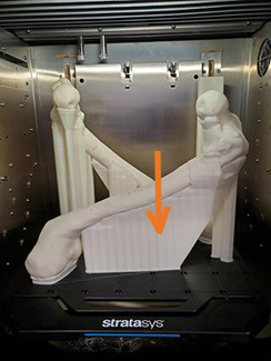 3D-printed models are composed of synthetic polymers, primarily plastic, and are printed with plastic filling the negative spaces of the models — like in between rib bones or spinal vertebrae. The orange arrow in the above photo shows negative space that is not needed for surgical planning and will be trimmed from a femur model after printing is complete.
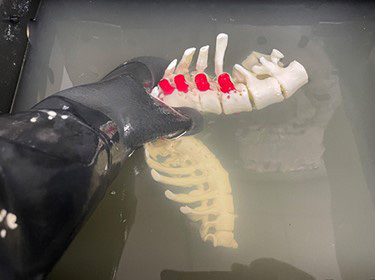 Instead of manually carving away the excess material in negative spaces, models are soaked in alkaline baths to dissolve the unneeded material. The red protrusions on the model shown above indicate where surgical hardware is present.
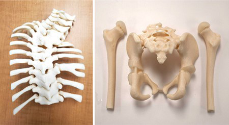 Final printed pieces: A curved spine of a scoliosis patient (left) and a pelvis with femurs (right).
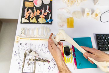
Not only do the printers build hard plastic materials but they also create squishy and rubbery parts, too. These parts mimic the feel and properties of actual tissue. In the photo above, elastic “cartilage” fits onto the head of a 3D-printed femur. This allows for a realistic connection between the bones and aids in surgical planning.
“Because CT scan images don’t show cartilage and other connective tissues, my team often partners with Radiology to cross-reference these parts with additional imaging, such as an ultrasound or MRI,” Dr. Friedman says. “Then, my team will engineer and incorporate these additional components into models so surgeons may accurately practice procedures and plan tools they will need.”
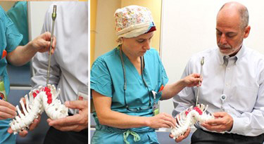
With a printed scoliosis model in hand, Dr. Bauer and Dr. Yaszay use surgical tools to determine how to best approach the patient’s condition and determine their plan for surgery.
National Leaders in Cutting-Edge Orthopedics and Sports Medicine Research
3D printing is just one of the cutting-edge technologies and research program that orthopedic patients can access at Seattle Children’s. The team conducts extensive research to improve treatments for children’s musculoskeletal conditions. Their studies focus on refining surgical techniques, testing new treatments, and enhancing patient outcomes through registries and clinical trials. Research areas include foot and ankle conditions, hip disorders, spine deformities, sports injuries, trauma, upper limb differences, and venous malformations. Collaborative efforts with national and international study groups help develop better treatment plans and rehabilitation strategies. By analyzing patient data and clinical outcomes, the team continuously advances care to ensure children lead active, healthy lives.
Refer a patient to Seattle Children’s Orthopedics and Sports Medicine or learn more about Seattle Children’s research programs.