White Matter Development and Injury
 A major cause of chronic disability in survivors of premature birth is diffuse white matter injury (DWMI) and hypomyelination. Altered development of the WM is directly associated with adverse outcomes, including cerebral palsy, cognitive delay and neurobehavioral problems.
The cellular pathophysiology underlying DWMI and abnormal myelination is complex and not fully understood. WM glia, and particularly oligodendrocytes (OLs) and their progenitors (OPCs), are susceptible to injury that often occurs in premature birth. We have previously used an animal model of hypoxia (HX)-induced global WMI to demonstrate that OPCs display delayed maturation, which results in abnormal myelination and altered WM function. We have uncovered major aspects of the cellular dysmaturation pathology underlying HX-induced delayed myelination in corpus callosum, including enhanced OPC proliferation associated with decreased OL differentiation, and disrupted myelin ultrastructure.
Recent analysis of OL development in corpus callosum (CC) demonstrates that:
- The prolonged proliferative state of OPCs and delayed OL differentiation in HX is a result of changes in HIF1α-dependent expression and activity of the histone deacetylases Sirt1 and Sirt2, respectively;
- HX reduces synaptic glutamate (Glu) release from cortical pyramidal neurons on OPCs, which normally downregulates OPC proliferation; and
- HX compromises axonal integrity and function in the subcortical white matter (SCWM), resulting in altered axon/myelin interactions.
Based on these results, we are now testing the hypothesis that HX-induced protracted WM immaturity arises from intrinsic (Sirt1 and Sirt2) and extrinsic (synaptic) dysregulation of OPC proliferation and OL maturation, thus affecting axonal integrity and function.
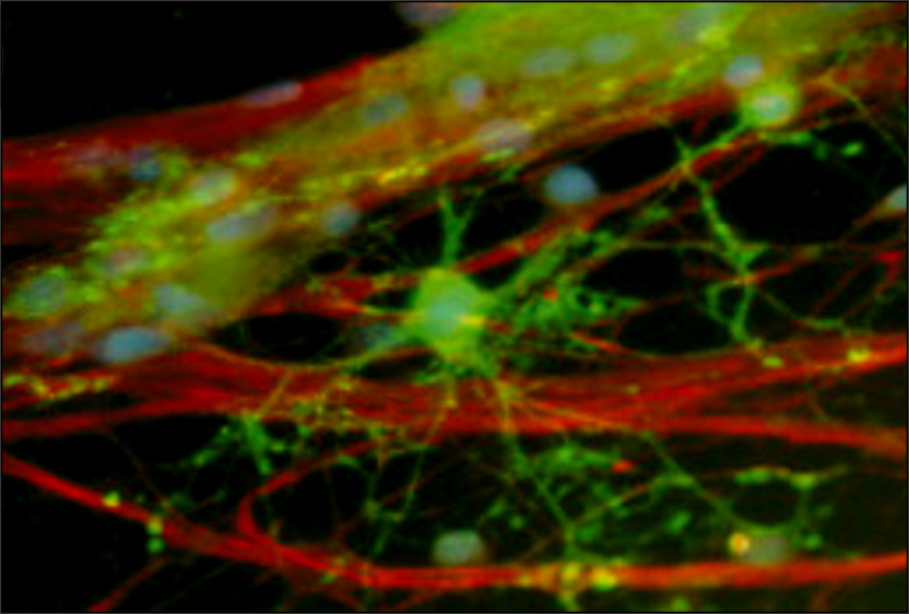
White matter oligodendrocytes. This image is mouse subcortical white matter oligodendrocytes and myelin in the CNP-EGFP mouse. Selective expression of green fluorescent protein in myelinating oligodendrocytes reveals multiple contacts between oligodendrocyte processes and axons.